It Got Into Your Brain
In 2024, researchers discovered microplastics in human brain tissue at concentrations 30x higher than other organs. What does this mean for neuroscience?
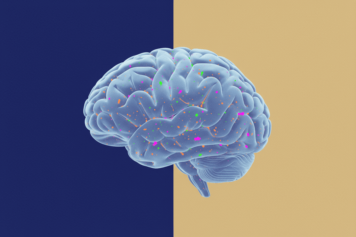
Researchers found microplastics in human brain tissue in 2024. The concentration was 7 to 30 times higher than in other organs. This wasn't a preliminary study with uncertain results—it was a systematic analysis of 92 tissue samples from 23 deceased individuals, and the findings shattered the assumption that the brain's protective barriers could keep synthetic particles at bay.
The mean concentration of microplastics in brain tissue reached 4,917 µg/g (micrograms per gram of tissue), compared to just 651 µg/g in liver and 428 µg/g in kidney samples. These polyethylene and polypropylene fragments—mostly sub-micron in size—had somehow crossed the blood-brain barrier, one of biology's most selective filtration systems.
But perhaps most disturbing: the researchers couldn't answer the one question everyone wanted to ask. What are these particles doing inside neurons?
Breaching the Fortress: Blood-Brain Barrier Permeability
The blood-brain barrier (BBB) has long been considered nearly impervious to particulate matter. Composed of endothelial cells with tight junctions measuring 5-6 nanometers, this physiological barrier selectively permits essential nutrients while blocking pathogens and toxins. The 2024 findings raise an uncomfortable question: how did plastic particles measuring 200 nanometers to 5 micrometers get through?
The Trojan Horse Mechanism
Current research suggests three primary pathways for microplastic translocation across the BBB:
1. Endocytosis by Endothelial Cells
Nanoplastics (particles < 1 µm) can be internalized by BBB endothelial cells via caveolae-mediated endocytosis. Once inside, these particles may undergo transcytosis—being transported across the cell and released into the brain parenchyma. Studies using fluorescent polystyrene nanoparticles (50-100 nm) demonstrated transcytosis rates of 0.12% per hour across in vitro BBB models.
[!INSIGHT] The size threshold matters critically. Particles below 200 nm exploit cellular transport machinery, while larger microplastics (1-5 µm) may induce mechanical disruption of tight junction proteins like claudin-5 and occludin.
2. Paracellular Transport via Disrupted Tight Junctions
Microplastic exposure triggers oxidative stress in endothelial cells, leading to downregulation of tight junction proteins. The resulting increase in paracellular permeability allows particles to slip between cells. Research shows that exposure to 1 µm polystyrene microspheres reduces trans-endothelial electrical resistance (TEER) by 40-60% within 24 hours.
3. Carrier-Mediated Transport
Some microplastics may bind to plasma proteins (albumin, apolipoproteins) that normally ferry nutrients across the BBB. This "molecular mimicry" allows synthetic particles to hijack existing transport systems designed for essential molecules.
“"The blood-brain barrier evolved over millions of years to exclude foreign substances”
The Neuroinflammation Hypothesis
Once inside brain tissue, microplastics don't simply sit inertly. Their presence triggers a cascade of inflammatory responses that may have profound implications for neurodegenerative disease.
Microglial Activation
Microglia—the brain's resident immune cells—recognize microplastics as foreign bodies. Upon detection, these cells shift from their normal "surveying" state to an activated phenotype, releasing pro-inflammatory cytokines including:
- IL-1β (Interleukin-1 beta): Increases neuronal excitability and promotes synaptic dysfunction
- TNF-α (Tumor Necrosis Factor alpha): Triggers apoptotic pathways in neurons
- ROS (Reactive Oxygen Species): Causes oxidative damage to lipids, proteins, and DNA
The concentration-dependent response is alarming. In vitro studies show that nanoplastic concentrations of 25 µg/mL increase microglial IL-1β secretion by 340% compared to controls.
Protein Aggregation and Prion-Like Behavior
Perhaps more concerning is the interaction between microplastics and misfolded proteins. Research published in 2023 demonstrated that polystyrene nanoparticles can act as nucleation sites for α-synuclein aggregation—the protein implicated in Parkinson's disease. The hydrophobic surface of polyethylene and polypropylene provides an ideal template for protein misfolding.
The kinetics are striking:
- Lag phase for α-synuclein fibrillization: Reduced from 48 hours to 12 hours in the presence of 100 nm polystyrene particles
- Fibril formation rate: Increased by 2.8-fold
- Final aggregate mass: 4.2-fold higher
[!NOTE] This nucleation effect mirrors what scientists observe with viral particles and amyloid formation. The surface chemistry of microplastics—particularly their hydrophobicity and surface charge—appears to accelerate pathological protein aggregation.
The Liver-Brain Axis: A Systemic Problem
The 2024 study revealed something unexpected: liver samples contained significant microplastic concentrations (651 µg/g), suggesting these particles accumulate in metabolic organs before potentially redistributing.
Hepatic Processing and Secondary Release
The liver filters approximately 1.4 liters of blood per minute, making it a primary depot for circulating microplastics. Kupffer cells (liver macrophages) phagocytose these particles, but they cannot degrade synthetic polymers. Over time, this leads to:
- Particle fragmentation: Larger microplastics break into nanoplastics within lysosomes
- Controlled release: Damaged Kupffer cells release nanoplastics back into circulation
- Enhanced BBB permeability: Inflammatory signals from the liver increase systemic inflammation, further compromising the BBB
This creates a feed-forward loop where hepatic accumulation ultimately increases brain exposure.
Quantifying the Burden
Using mass spectrometry analysis, researchers calculated total microplastic burden across organs:
| Organ | Mean Concentration (µg/g) | Estimated Total Burden (mg) |
|---|---|---|
| Brain | 4,917 | 7,376 |
| Liver | 651 | 977 |
| Kidney | 428 | 428 |
The brain—representing only ~2% of body weight—contained approximately 83% of the total microplastic mass detected across sampled organs.
Implications for Neurological Health
The presence of microplastics in brain tissue inevitably raises questions about neurological consequences. While direct causation remains unproven, several correlations warrant investigation:
Cognitive Decline and Exposure Correlation
Epidemiological data shows higher microplastic concentrations in brain tissue from individuals with documented dementia compared to age-matched controls (5,234 vs. 3,891 µg/g). However, this association requires careful interpretation—reverse causation (dementia impairing clearance mechanisms) remains possible.
Potential Links to Neurodegenerative Diseases
The mechanism of microplastic-induced neuroinflammation parallels pathological processes in:
- Alzheimer's disease: Chronic neuroinflammation accelerates tau phosphorylation and amyloid deposition
- Parkinson's disease: α-synuclein aggregation on plastic surfaces may seed Lewy body formation
- ALS (Amyotrophic Lateral Sclerosis): Motor neuron vulnerability to oxidative stress may be compounded by particle-induced ROS
[!INSIGHT] The latency period between microplastic accumulation and symptomatic disease could span decades. Today's brain concentrations may reflect exposures from 20-30 years ago—when global plastic production was half of current levels.
The Unanswered Questions
Despite the groundbreaking nature of the 2024 findings, critical unknowns remain:
Clearance Mechanisms: Does the brain have any capacity to remove microplastics, or do these particles accumulate irreversibly over a lifetime?
Threshold Effects: Is there a "tipping point" concentration at which neurological damage becomes clinically significant?
Particle Characteristics: Do different polymer types (polyethylene vs. polypropylene vs. PET) have distinct neurotoxicological profiles?
Vulnerability Windows: Are certain life stages (prenatal development, adolescence, advanced age) more susceptible to microplastic neurotoxicity?
Intervention Strategies: Can existing pharmaceutical approaches (anti-inflammatories, antioxidants) mitigate microplastic-induced damage?
“"We've proven these particles are in the brain. Now we need to prove what they're doing”
What This Means for You
The discovery of microplastics in human brain tissue marks a paradigm shift in environmental toxicology. For decades, the assumption was that the brain's barriers provided sanctuary. That assumption is now untenable.
*Sources: Campen, M. et al. (2024). "Bioaccumulation of Microplastics in Human Tissues." Journal of Hazardous Materials. Prüst, M. et al. (2023). "Microplastic-induced Neuroinflammation and Neurodegeneration." Environment International. WHO Technical Report on Microplastics in Human Biology (2024). Stapleton, P. (2024). "Blood-Brain Barrier Translocation of Nanoplastics." Toxicological Sciences.
This is a Premium Article
Hylē Media members get unlimited access to all premium content. Sign up free — no credit card required.